A dental x-ray is one of the procedures patients question most. When the dentist says "let's take a film," the same thoughts tend to surface: Is this really necessary? Isn't radiation harmful? How often can I have one? The hesitation is understandable. But when a dental x-ray is involved, the useful question is not "is …
A dental x-ray is one of the procedures patients question most. When the dentist says “let’s take a film,” the same thoughts tend to surface: Is this really necessary? Isn’t radiation harmful? How often can I have one?
The hesitation is understandable. But when a dental x-ray is involved, the useful question is not “is there any radiation?” — it is “how much, and what do we gain in return?” A visual examination reveals only the visible surface of a tooth. An infection inside the bone, decay starting between two teeth, or an impacted wisdom tooth simply cannot be seen with the naked eye.
In this article, the dentists at Bilim Dental Clinic in Maltepe, Istanbul explain what each imaging method is for, how much radiation a panoramic film actually involves, what safety measures apply during pregnancy and childhood, and the questions patients ask us most.
Why Is a Dental X-Ray Taken?
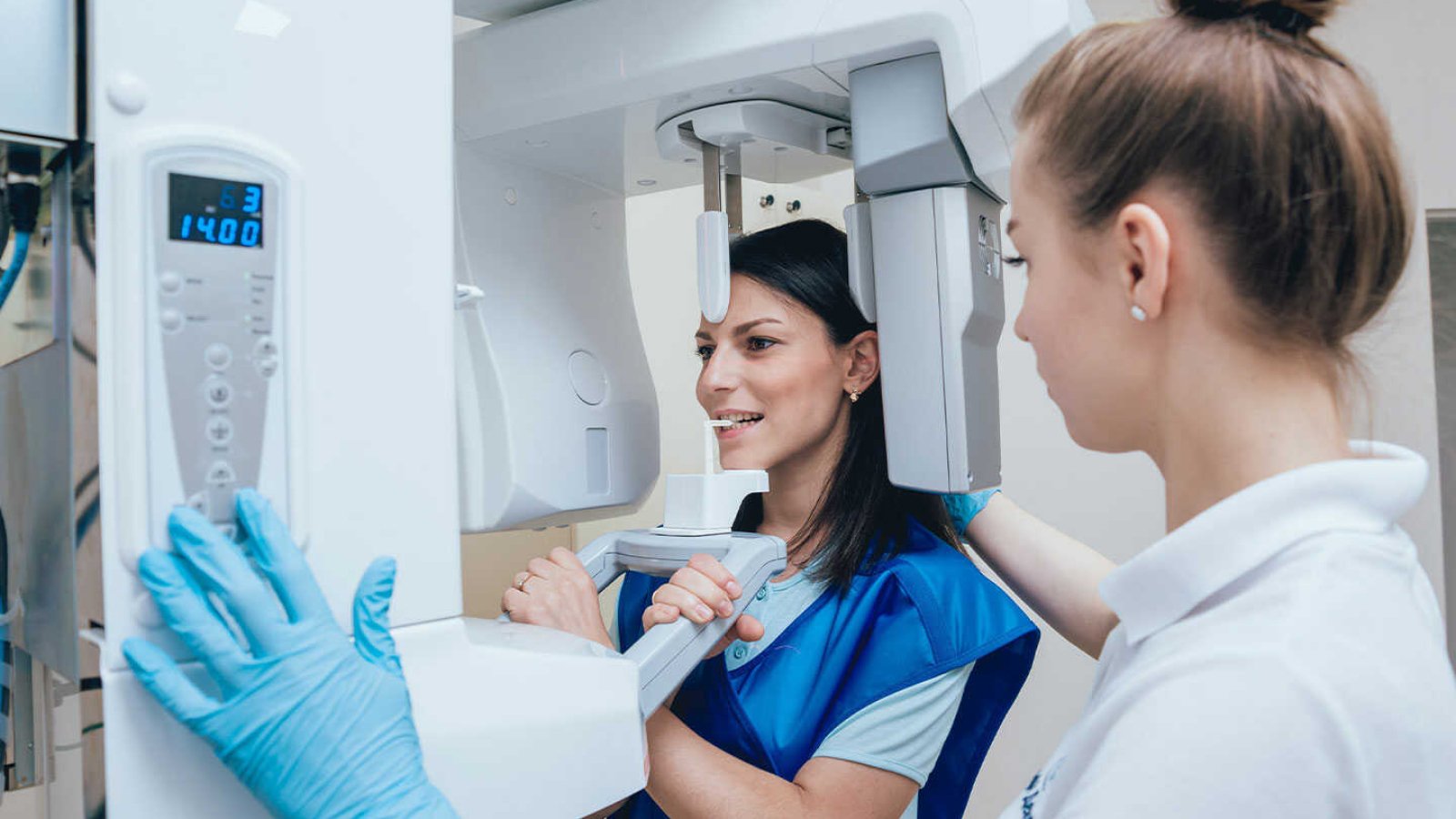
During a clinical examination, the dentist can inspect only the part of the tooth visible above the gum. A significant portion of every tooth sits inside gum and bone. A dental x-ray makes that hidden region visible.
The most common reasons include:
- Decay that has begun between teeth and cannot be seen directly
- Infection or cysts at the root tip
- Measuring bone levels to determine the severity of gum disease
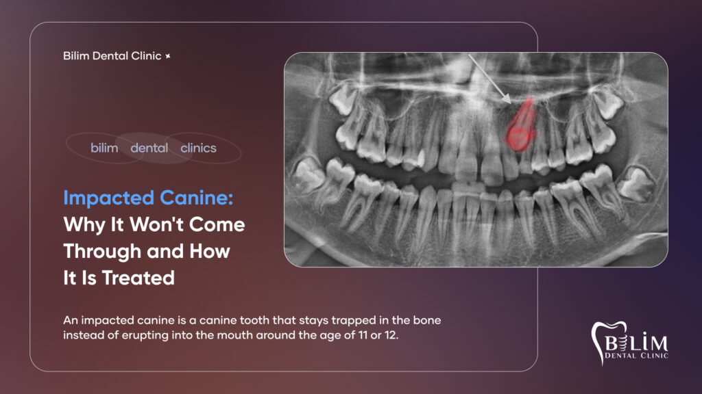
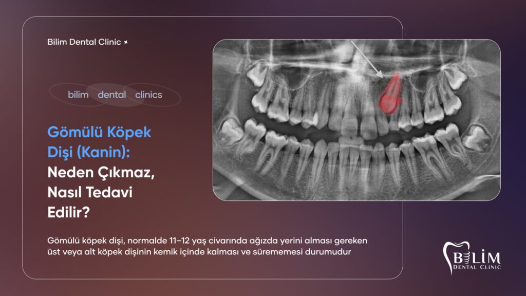
- The position of impacted teeth and their relationship to nearby structures
- The development of permanent teeth beneath the baby teeth
- Checking the result after a filling, crown, or root canal
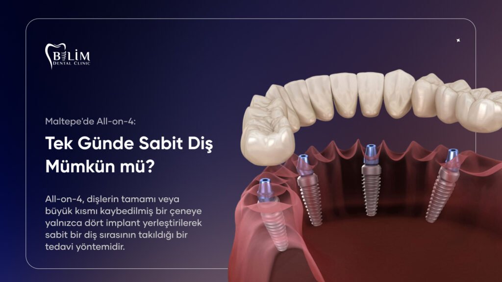
- Measuring bone height and width before implant placement
Without this information, a treatment plan is incomplete. An implant placed without measuring the available bone, or root canal treatment started without knowing how many canals a tooth has, carries a real risk of problems later.
Types of Dental X-Ray and What Each One Shows
In everyday conversation every dental x-ray is called a “film”, but each type answers a different question.
| Method | What It Shows | Average Dose (μSv) | When It Is Used |
|---|---|---|---|
| Periapical film | One tooth in full, including the root tip | 1–8 | Root canal treatment, root-tip infection, single-tooth complaints |
| Bitewing | The contact surfaces of the back teeth | 1–8 | Early detection of decay between teeth |
| Panoramic film | Both jaws, all teeth, sinuses, jaw joints | 14–24 | General assessment, impacted teeth, first examination |
| Cephalometric film | Profile view of the skull and jaw relationship | 3–6 | Orthodontic treatment planning |
| CBCT (cone beam CT) | Three-dimensional bone and anatomy | 20–200 | Implant planning, complex surgical cases |
The guiding principle is simple: answer the smallest question with the smallest method. If one tooth hurts, a panoramic film is unnecessary — a single periapical film will do.
Periapical and Bitewing Films
These are taken with a small sensor placed inside the mouth. A periapical film shows a single tooth from crown to root tip, which is what you need for a painful tooth, for following root canal treatment, or when infection at the root tip is suspected. Bitewings show where the back teeth touch each other, making them the most effective method for catching early decay.
Panoramic Films
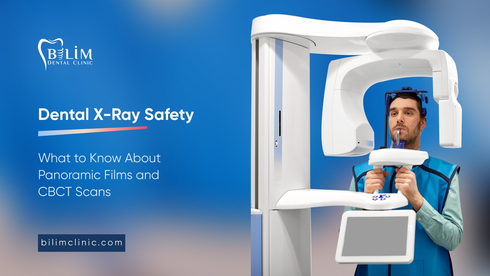
This is a single image of the entire mouth, captured by a machine that rotates around the head. It provides a map: all the teeth, the jawbones, the floor of the sinuses, the jaw joints, and any impacted teeth, all at once. It is the usual choice at a first examination or when planning comprehensive treatment. The proximity of an impacted wisdom tooth to the nerve canal is also first assessed here, and that information shapes how oral and maxillofacial surgery is planned.
Panoramic films do have a limitation: they are two-dimensional, so structures can overlap. That is why they are less reliable than bitewings for spotting small areas of decay between teeth.
Cephalometric Films
This profile image measures the position of the upper and lower jaws relative to each other and to the base of the skull. It is used in orthodontic treatment planning, in assessing the direction of growth, and for comparison once treatment is complete.
CBCT (Cone Beam Computed Tomography)
CBCT produces a three-dimensional image of the jaw region. It differs from medical CT: it scans only the jaws, and the dose is considerably lower. It measures bone thickness, the path of the nerve canal, and the sinus floor to within a millimetre, which is why it has become a standard part of planning implant treatment.
How Much Radiation Does a Dental X-Ray Involve?
This is the question everyone really wants answered. The clearest way to make the numbers meaningful is to compare them with the natural background radiation we are all exposed to anyway.
The global average background exposure is roughly 2,400 μSv per year — about 6 to 10 μSv per day. It comes from the ground, the air, our food, and outer space, and it cannot be avoided.
On that scale:
- Periapical or bitewing film (digital): roughly 1–8 μSv → less than a single day of background radiation
- Panoramic film: roughly 14–24 μSv → about two to three days of background radiation
- CBCT scan: roughly 20–200 μSv → from a few days to about a week, depending on the field size
- For comparison, a chest x-ray: roughly 100 μSv
- For comparison, a long-haul flight: roughly 30–80 μSv
In other words, a panoramic film usually involves less exposure than a flight from Istanbul to Western Europe. That does not mean radiation is trivial — every unnecessary exposure is avoided. But when a film is genuinely needed, its risk is far smaller than the risk of an undiagnosed infection or an unnoticed cavity.
Dentistry follows a principle known as ALARA: keep the dose As Low As Reasonably Achievable. In practice this means not taking a dental x-ray without a reason, keeping the field as small as possible, and using modern digital equipment.
Is a Lead Apron Still Necessary for a Dental X-Ray?
For many years the lead apron was standard. More recently, international radiology organisations have revised their guidance: in modern machines the beam is tightly collimated, so it does not reach the area the apron covers in the first place. In some cases an apron can even interfere with a machine’s automatic exposure control and cause the image to be repeated.
Even so, many clinics continue to use aprons because patients find them reassuring and the practice is well established. Thyroid collars remain in widespread use, particularly for children and for images taken close to the beam. Feel free to ask which approach your clinic follows — both are safe.
What Happens During a Panoramic Film?
The procedure takes about 15 to 20 seconds and is completely painless. The only thing asked of you is to stay still.
First, metal jewellery, glasses, hair clips, and any removable denture are taken off, since these cast shadows on the image. You then rest your chin on the support and bite gently on a positioning stick. You will be asked to press your tongue against the roof of your mouth, which prevents an air shadow from forming on the image. The machine slowly rotates around your head while you remain motionless. The most common problem is a small movement mid-scan, which can mean repeating the film.
For anyone concerned about enclosed spaces, one reassurance: the machine is open, you stand upright, and nothing surrounds you.
Does Every Patient Need a CBCT Scan?
No. CBCT is reserved for situations where a two-dimensional image is not enough. It is not requested for a routine check-up or a simple filling.
The typical indications are implant planning, clarifying the relationship between an impacted tooth and the nerve canal, suspected cysts or tumours in the jaw, jaw joint problems, complex root canal anatomy, and orthognathic surgery planning.
When a scan is suggested, the most useful question a patient can ask is: “How will this image change my treatment plan?” Any request with a clear answer to that question is a justified one.
Can You Have a Dental X-Ray During Pregnancy?

This is the question that causes the most anxiety. The consensus among scientific bodies is that imaging can be carried out when it is necessary, with appropriate precautions. The beam is directed at the jaw, far from the abdomen, and scattered dose from modern equipment is extremely low.
Practice nevertheless remains cautious. Non-urgent imaging is postponed until after delivery. Where delay carries the greater risk — severe pain, an abscess, or spreading infection — the film is taken, because an untreated dental infection poses a far greater danger to mother and baby than the imaging does.
Always tell your dentist if you are pregnant or think you might be. Our article on dental care during pregnancy covers this period in more detail.
Imaging in Children
Children are more sensitive to radiation than adults, because their tissues divide rapidly and they have a long life ahead of them. For that reason, the decision to take a dental x-ray in a child is made even more carefully.
Precautions include reducing the exposure setting to suit the child’s size, narrowing the field, and taking as few images as possible. Imaging is still sometimes unavoidable: monitoring the development of permanent teeth, checking for a missing tooth bud, assessment after trauma, and orthodontic planning are the main reasons. The decision always rests on weighing benefit against risk.
What the Dentist Actually Reads on a Dental X-Ray
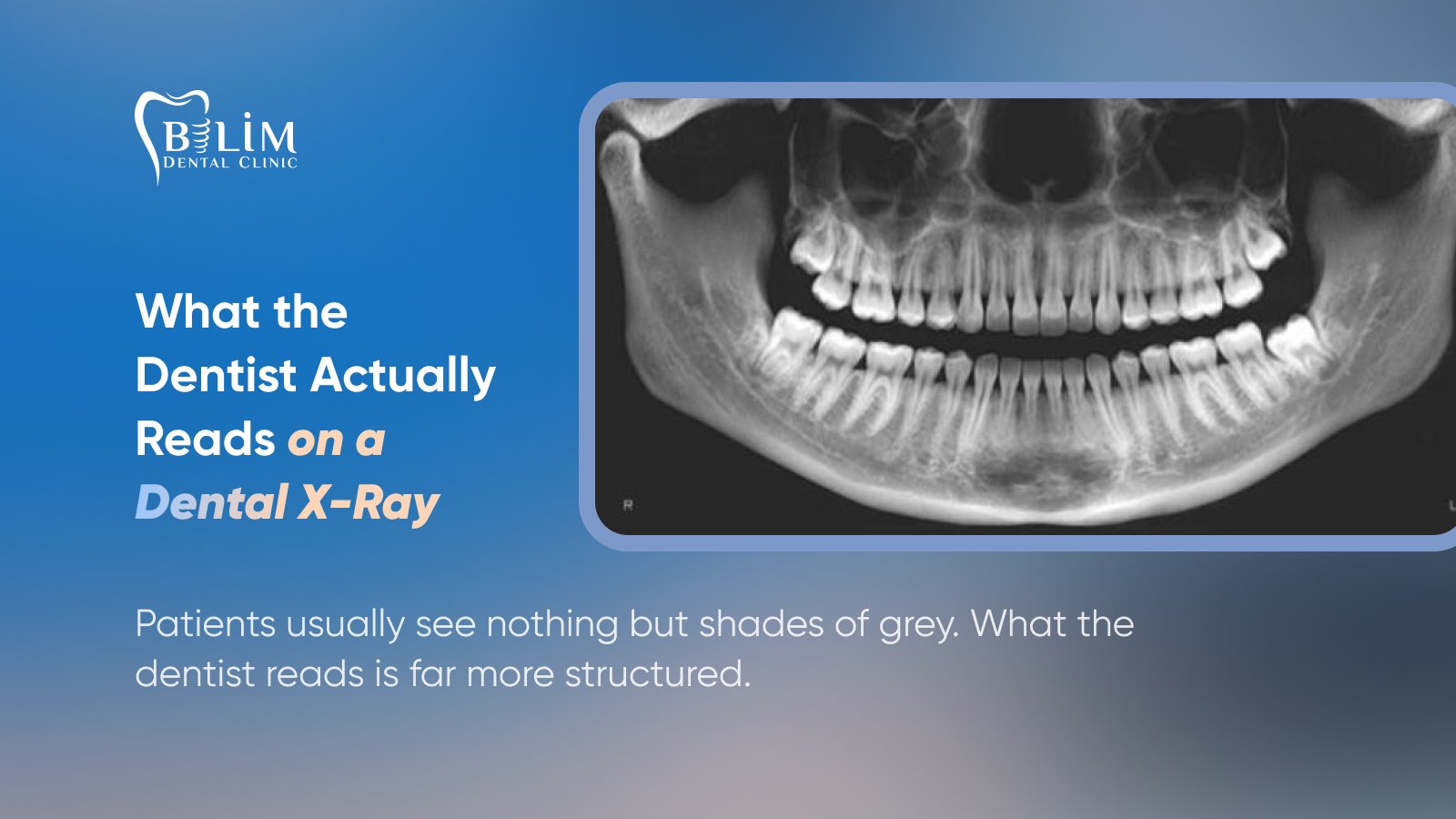 Patients usually see nothing but shades of grey. What the dentist reads is far more structured.
The first thing checked is the number and position of the teeth: is anything missing, is a tooth impacted, which way do the roots point. Next come the dark areas between teeth and around the edges of fillings, which suggest decay — decayed tissue absorbs less radiation than healthy tooth, so it appears darker.
Then comes the bone. The level of the bone surrounding each tooth is measured against the length of the root. When that level drops, gum disease has reached the bone. Dark, rounded areas at the root tips indicate chronic infection, and these frequently appear in patients who have no pain at all.
Finally, the anatomy is reviewed: the sinus floors, the path of the nerve canal in the lower jaw, the appearance of the jaw joints. A dental x-ray sometimes reveals findings unrelated to the teeth — signs of sinus inflammation or structural changes in the jawbone are occasionally noticed here first.
Imaging for International Patients
For patients travelling to Turkey for treatment, a dental x-ray is the first step of planning. Many send a panoramic film taken in their own country beforehand, which is often enough for an initial assessment and a draft treatment plan.
The definitive plan, however, is confirmed with current imaging once the patient arrives. Films vary in quality, and the situation may have changed in the meantime. Where implants are planned, a three-dimensional scan is almost always required.
If you are sending images ahead of your visit, share the original digital file — ideally in DICOM format — rather than a screenshot, and always include the date it was taken. You can find more about how we organise treatment for visiting patients on our dental tourism page.
Common Myths
“If I’m not in pain, I don’t need a film.” False. Root-tip infections and decay between teeth often progress without pain. By the time symptoms appear, the situation is usually more advanced.
“Radiation accumulates in the body.” False. The radiation used in diagnostic imaging is not stored in the body. Exposure is limited to the moment of the scan.
“I’m pregnant, so it’s absolutely forbidden.” False. Imaging can be performed when needed, with appropriate precautions. The greater risk is leaving an infection untreated.
“A panoramic film shows everything.” False. It gives a general map; small cavities and three-dimensional measurements require other methods.
“Digital and traditional films are the same.” False. Digital systems produce the same image at a noticeably lower dose.
“I had one once, so it’s valid forever.” False. An image shows the situation on the day it was taken. Teeth and bone both change over the years.
How Often Should a Dental X-Ray Be Taken?
There is no single interval that suits everyone. Frequency depends on your risk of decay and your dental history.
An adult with a low risk of decay, healthy gums, and regular check-ups may need a review film only every few years. For someone who develops decay frequently, has advanced gum disease, or is in active treatment, the interval shortens. Images taken during treatment are separate from this: several small films during root canal treatment, for example, are entirely normal.
One important reminder: a dental x-ray is not taken by the calendar. Every image should have a clinical reason behind it.
Keeping Copies of Your Images
Every dental x-ray is a record of a moment, and it becomes more valuable with time. A single image shows your current situation; two images show change — and change is where the real information lies.
If you switch dentists, move city, or want a second opinion, having your previous records serves two purposes: the new dentist sees the history, and an unnecessary repeat scan is avoided. Do not hesitate to ask your clinic for a digital copy of your images; it is entirely your right as a patient.
Name the files with the date they were taken. A folder on your phone is enough for most people. Clinics keep records for the period the regulations require, but having your own copy makes life much easier if you are treated at more than one practice.
What Digital Systems Changed
The shift from chemically developed film to digital sensors is one of the most significant improvements for patients. Digital systems produce the same image at a markedly lower dose. The image also appears instantly on screen, can be magnified, has adjustable contrast, and can be stored for comparison with future images.
That comparison is more valuable than it sounds. Place two films taken two years apart side by side and a slow loss of bone or an advancing cavity becomes obvious — where either image alone would look unremarkable.
Frequently Asked Questions
Am I obliged to have a dental x-ray?
No, not every treatment requires imaging. But if your dentist requests one, there is a clinical reason behind it. Feel free to ask what that reason is; no image should be taken without a justification you can understand.
Is it a problem to have several films on the same day?
The combined dose of a few small films plus a panoramic image remains at the level of a few days of background radiation. If there is a clinical reason, it is not a concern.
Can images be taken while breastfeeding?
Yes. Dental imaging does not affect milk production or the milk itself. There is no need to interrupt breastfeeding.
I have a thyroid condition — can I still have imaging?
Yes. You can request that a thyroid collar be used if it makes you more comfortable. Simply tell your dentist about your condition.
Does a dental x-ray feel like anything?
No. The beam cannot be seen, heard, or felt. The only discomfort is a brief gag sensation from the sensor touching the palate during intraoral films.
Does a panoramic film show cavities?
It shows larger cavities, but bitewings are the better choice for small, early decay between teeth.
Can I bring old images to a new dentist?
Yes, and you should. Previous images allow comparison and often prevent an unnecessary repeat.
Is CBCT always necessary for implants?
In most cases, yes. Bone thickness and the position of the nerve canal can only be measured reliably in three dimensions. Some straightforward cases may be planned with a panoramic film; your dentist decides.
Do metal fillings or crowns spoil the image?
They create bright areas, but this rarely prevents the dentist from assessing the region. The effect can be more noticeable on CBCT scans.
Can I stay with my child during their scan?
Usually yes, and you will be given protective covering. It depends on the individual clinic’s policy.
Dental Imaging at Bilim Dental Clinic in Maltepe
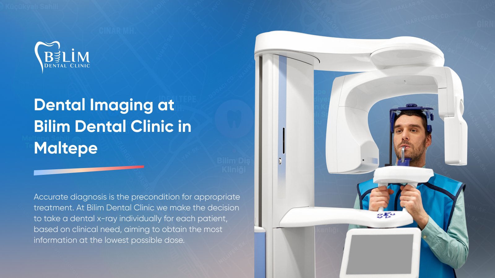
Accurate diagnosis is the precondition for appropriate treatment. At Bilim Dental Clinic we make the decision to take a dental x-ray individually for each patient, based on clinical need, aiming to obtain the most information at the lowest possible dose. In this article we have gathered the questions our patients ask most: what each type of film is for, how much radiation is involved, how we approach pregnancy and childhood, and how often imaging is appropriate.
At our clinic, we provide services in all areas of oral and dental health — from aesthetic dentistry and implant treatments to orthodontics and prosthetic applications — using modern technology and evidence-based, scientific approaches. Our experienced team of dentists creates a personalized treatment plan for every patient, aiming to build healthy, natural, and aesthetic smiles.
The dentists who will accompany you throughout this process:
- Dt. Ali Enes Öztürk
- Dt. Salman Taştan
- Dt. Hüseyin Kaya
- Uzm. Dt. Burak Güleç (Orthodontics Specialist)
- Dt. Cem Karaman
- Dt. Ayşe Sizer Çakar
- Dt. Zeynep Ece Okur (Pediatric Dentist)
For an examination, imaging, or treatment planning, we invite you to visit us on Bağdat Caddesi in Maltepe, Istanbul. You can follow our latest updates on our Instagram account.
WhatsApp: +90 538 977 89 01